15 + Corona Radiata Axial Mri High Quality Images. Inferiorly these tracts converge as the internal capsule. Analyzing the corona radiata, axial diffusivity (AD) and MD were significantly increased in the left superior region, MD and RD were Request Full Text Paper.

21 + Corona Radiata Axial Mri HD Wallpapers
Based on this output, the other ROA placements will be clearer.

View Image

Corona radiata


ECR 2015 / C-0510 / The value of MRI Brain following ...
Lentiform Nucleus Anatomy Ct - takvim kalender HD

Permanent brain MRI injury after electrical shock | Eurorad
Gehirn: MRT-Atlas der menschlichen Anatomie

Diffusion-Weighted Magnetic Resonance Imaging Identifies ...

Normal Anatomy | Radiology Key

Permanent brain MRI injury after electrical shock | Eurorad

Structural MRI results. From left to right: T1-3D sagittal ...
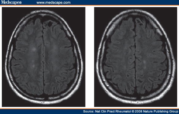
Optic Neuritis and Recurrent Myelitis in Patient With SLE
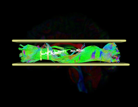
Anterior Corona Radiata – acr | Whole Brain Protocol for ...

MRI, CT findings in ALL with leptomeningeal disease
Cureus | Acute Post-stroke Hemiparkinsonism and ...
Axial T2-weighted magnetic resonance imaging (MRI) of t ...
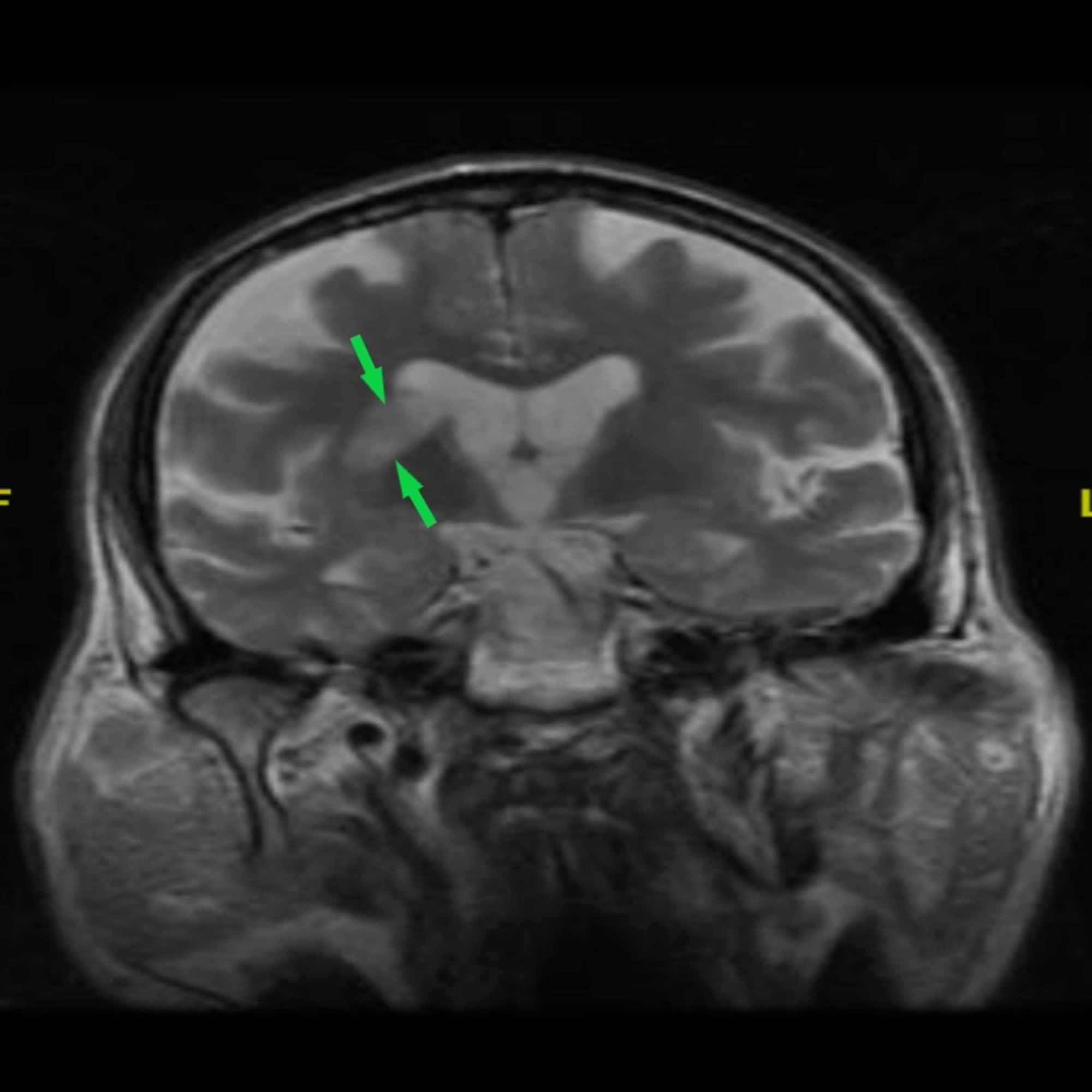
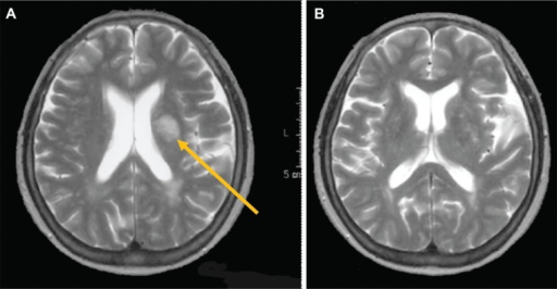
15 + Corona Radiata Axial Mri Desktop WallpaperRestricted corona radiata infarcts can occur near the lateral angle of the lateral ventricles. Arrows point to the major pathological features. from publication: Two cases. Please type a message to the paper's authors to explain your need for the paper.